Structural Characterization of Some Medicinal Weeds (Portulaca oleracea, Melissa officinalis and Peperomia pellucida) at Rivers State University
-
Ajuru Mercy Gospel


Department of Plant Science and Biotechnology, Rivers State University, Nkpolu-Oroworuokwo, Para Medical Board 5080, Port Harcourt, Rivers, Nigeria
Favour WorluDepartment of Plant Science and Biotechnology, Rivers State University, Nkpolu-Oroworuokwo, Para Medical Board 5080, Port Harcourt, Rivers, Nigeria
| Received 19 Jan, 2023 |
Accepted 17 Jun, 2023 |
Published 30 Sep, 2023 |
Background and Objectives: Melissa officinalis is a member of Lamiaceae family that is said to possess antifungal and antiviral properties, while Peperomia pellucida belongs to the family Piperaceae and is found in most African countries which can be used to reduce glucose and uric acid levels. Lastly, Portulaca oleracea is an annual plant that belongs to the family Portulacaceae which is used as a common vegetable. This study investigated the anatomical and histochemical structures of the stem and petiole of Melissa officinalis L. (lemon balm) Peperomia pellucida L. (pepper elder) and Portulaca oleracea L. (common purslane) in Rivers State University. This investigation aimed toprovide basic anatomical and histochemical data that will characterize and distinguish among the three species. Materials and Methods: Plant materials were identified, collected and fixed in formaldehyde: Acetic acid and alcohol (FAA) solution for three days. The hand-sectioning method was employed for both studies, in addition to the application of hydrogen peroxide and silver nitrate for 30 min in the histochemical study. Results: The anatomical structures are relatable due to their similarities in the layer of the epidermis and vascular bundles. The histochemical results showed that they possess calcium oxalate crystals apart from Peperomia pellucida . Conclusion: The standardized anatomical characters assisted in the detection and diagnosis of particular medicinal plant species.
| Copyright © 2023 Gospel and Worlu. This is an open-access article distributed under the Creative Commons Attribution License, which permits unrestricted use, distribution, and reproduction in any medium, provided the original work is properly cited. |
INTRODUCTION
Ever since prehistoric times, medicinal plants have been discovered and used in traditional medicine practices. Hundreds of chemical and biochemical compounds are synthesized by plants for functions such as defense against insects, herbivorous mammals, fungi and diseases. A lot of phytochemicals with potential biological activity have been discovered in plants and a single plant is discovered to contain diverse phytochemicals, leading to the uncertainty of using a whole plant as a medicine. Further, since the pharmacological actions and phytochemical contents of several plants with medicinal potential remain untapped and unassessed by rigorous scientific research, then it is difficult to categorically define safety and efficacy1.
According to Ward et al.2 a weed is defined as “a plant that forms populations that can enter habitats cultivated, markedly disturbed or occupied by man and potentially depress or displace the resident plant populations which are deliberately cultivated or are of ecological and/or aesthetic interest.”
Weeds are used as fodder, vegetables, manure, fuel, mats and screens, ornamental flowers, fencing purposes, soil binders, indicator plants and food for wildlife3,4.
Peperomia pellucida is a plant mostly found in humid areas and has a height of 6-45 cm with an erect stem, though, sometimes striated and epiphytic. The leaves have a width of about 0.3-4 cm. The petiole is round with longitudinal furrows of about 0.1-0.7 cm long, fleshy, oval and triangular. Leaves have wide bases which are conical at the ends with a whole edge. There are 3-5 leaf veins at the base of the flower which grow upwards in the opposite direction, with a length of about 0.8-9.2 cm. The chemical constituents from these plant extracts include alkaloids, flavonoids, saponins, tannins and triterpenoids. The plant is used in treating ailments such as burns and heat, kidney disease, headaches and fever.
Portulaca oleracea, commonly called little hogweed, is an annual or perennial plant that grows in the tropics and belongs to the family Portulacaceae. The stems are glabrous in nature, fleshy, purplish-red to green, originating from a taproot, often prostrate, forming mats. The leaves are also fleshy, alternately arranged on stems, though sometimes sub-alternate or opposite, obovate to spatulate with an apex that is obtuse or truncate-emarginate. The leaves size may be from 40×15 up to 60×25 mm when grown in fertile soils. There are little or no axillary hairs. Flowers are borne in a group at the end of the stem5. The petals are 4-6 in number, yellow in colour and range from 3 to 10 mm long by 2 to 8 mm wide with 6-15 stamens. The seeds are red or brown but black when mature, 0.6-1 mm long, usually with granules to flat-stellate surfaces.
Melissa officinalis is commonly called lemon balm, bee balm and honey balm. It is a perennial herb and belongs to the Lamiaceae family. The plant belongs to a genus that includes 5 species of perennial herbs native to Europe, Central Asia and Iran. Although Melissa officinalis originated primarily in Southern Europe, it is now naturalized around the world, from North America to New Zealand.
Lemon balm is commonly used as a sedative and tranquillizer and as an anti-gas. It reduces fever, it has antibacterial, spasmolytic, hypotensive, memory-enhancing, menstrual-inducing and thyroid-related effects, it has antiviral, antioxidant, antifungal, antiparasitic and antispasmolytic activities, it is also used to treat flatulence, asthma, bronchitis, amenorrhea, cardiac failure, arrhythmias, ulcers and wounds6.
There is a dearth of information on the anatomical and histochemical structures of the three plants studied: Melissa officinalis, Portulaca oleracea and Peperomia pellucida. Therefore, this work is aimed at characterizing the anatomical and histochemical structures of the three plants studied: Melissa officinalis, Portulaca oleracea and Peperomia pellucida at Rivers State University.
MATERIALS AND METHODS
Plant collection, identification and preparation: The study was carried out from July, 2022 to November, 2022. The three plant materials for investigation (Portulaca oleracea, Melissa officinalis and Peperomia pellucida) were collected from the botanical garden at Rivers State University and were taken to the Department of Plant Science and Biotechnology Laboratory at Rivers State University. The parts of the plants used were the stem and petiole. The plants were identified and authenticated by a plant taxonomist in the Department. The plants were properly dried and pressed and the voucher specimens were deposited in the University’s Herbarium.
Anatomical study: Specimen bottles were gotten and labelled accordingly and plant materials were prepared by cutting them into small sections and placing them in the specimen bottle and each specimen bottle was filled with FAA (Formalin: Acetic acid: 95% Alcohol) in a ratio of 1:1:18, respectively. Plants were removed from the FAA mixture. The plant materials were rinsed in three changes of distilled water with Petri dishes already filled with distilled water.
Sections of the stem and petiole for each plant were made, while a camel hairbrush was used to lift the sections from the Petri dishes unto the slides and excess water was removed from the slide using filter paper. A drop or two of 1% safranin was placed on the sections and the setups were kept for 5 min after which distilled water was used to rinse off excess safranin and filter paper was used to remove excess water. Then a drop or two of glycerin were placed on the setups while filter papers were used again to remove the excess mountant after which coverslips were used to cover the sections to avoid trapping air bubbles and finally, after 1 week, BS-2025T binoculars microscopes by Bestcope were used to view the slides.
Histochemical study: Specimen bottles were gotten and labelled accordingly and plant materials were prepared by cutting them into small sections and were placed in a specimen bottle and each specimen bottle was filled with a Formalin-Acetic acid-Alcohol (FAA) mixture.
The plant materials were rinsed in three changes of distilled water with a Petri dish already filled with distilled water. The sections of plant materials were cut, stem and petiole, while a camel hair brush was used to lift the sections from the Petri dishes onto a slide and excess water was removed using filter paper. The slides containing the sections were placed under high current light and a drop or two of hydrogen peroxide was placed on the sections and excess of it was removed immediately. Then a drop or two of silver nitrate was placed on the sections and allowed to stay for 15-20 min and again excess of it was removed using filter paper. Then a drop or two of glycerin was placed on the setups while filter paper was used again to remove the excess glycerin after which coverslips were used to cover the sections and avoid trapping air bubbles finally after 1 week the slide was viewed under the BS-2025T binocular microscopes by Bestcopes microscope. Parameters determined were calcium oxalate crystals present, the tissue(s) found and the frequency of appearance.
RESULTS
Anatomical study
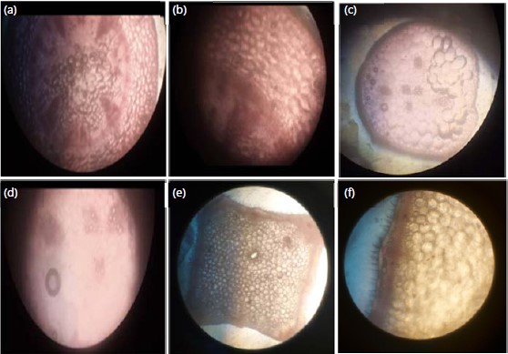
Stem anatomy: The results of the stem anatomical structures of the plants studied (Melissa officinalis, Peperomia pellucida and Portulaca oleracea) were presented in Fig. 1 and Table 1.
| Table 1: | Stem anatomical characteristics of the plants studied | |||
| Parameters | Melissa officinalis | Portulaca oleracea | Peperomia pellucida |
| Stem anatomy | |||
| Epidermis | 1 layer | 1 layer | 1 layer |
| Collenchyma | 2-3 layers | 2-3 layers | 3-4 layers |
| Parenchyma | 2-3 layers | 7-8 layers | 3-4 layers |
| Endodermis | 1 layer | 1 layer | 1 layer |
| Pericycle | 1 layer | 1 layer | 1 layer |
| Vascular bundle | |||
| a: Number | They are numerous. | There are 15 vascular bundles | The vascular bundle are not arranged |
| b: Arrangement | The vascular bundles are collateral, conjoint and open |
The vascular bundle is arranged in a ring. They are collateral, conjoined and open |
in a ring, but they are conjoined and open |
| Pith cavity | The pith cavity is filled with parenchyma cells |
The pith cavity is filled with parenchyma cells |
The pith cavity is filled with parenchyma cells |
|
The stem of Melissa officinalis is square-shaped and the epidermal layer is one-celled-thick, covered by the cuticle. There are numerous glandular multi-layered and multiseriate trichomes. The epidermal layer is closely followed by two to three layers of collenchyma cells. Beneath the collenchyma cells are two to three layers of parenchyma cells. The endodermis, which follows the parenchyma, is one layer thick. A uniseriate pericycle surrounds the vascular bundles arranged on the sides of the square-shaped stem. The pith cavity is very wide filled with parenchyma cells with large intercellular spaces.
In Peperomia pellucida, the epidermal cell is one layer thick and covered by a thick cuticle, it is followed by three to four layers of collenchyma cells, followed by three to four layers of parenchyma cells with intercellular air spaces. The endodermis surrounds the pericycle and they are both one layer thick. The vascular bundles are arranged in a ring and they are conjoined and open. The xylem vessels are towards the center while the phloem is towards the periphery in the pith cavity which is filled with parenchyma cells.
In Portulaca oleracea, the epidermal layer is one-celled thick and covered by a thin cuticle, there are no trichomes. Below the epidermis, there are two to three layers of collenchyma cells followed by seven to eight layers of parenchyma cells with intercellular air spaces. A layer of endodermis surrounds the pericycle which is also one layer thick. The vascular bundles are arranged in a ring. They are collateral, conjoined and opened. They are 15 vascular bundles: The xylem cells are towards the center while the phloem cells are towards the periphery. The pith cavity is filled with parenchyma cells with intercellular air spaces.
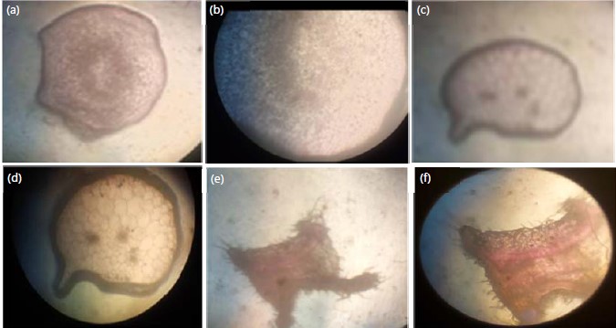
Petiole anatomy: The results of the petiole anatomical structures of the plants studied (Melissa officinalis, Peperomia pellucida and Portulaca oleracea) were presented in Fig. 2 and Table 2.
The anatomical structure of the petiole of Melissa officinalis showed that the epidermal layer is one-celled thick, covered by a thick cuticle. There are numerous glandular multi-cellular trichomes all over the epidermis. The epidermis is followed by the cortex which is two-three layers thick. There are 2 vascular bundles with parenchyma cells having intercellular spaces filling the pith cavity.
|
| Table 2: | Petiole anatomical characteristics of the plants studied | |||
| Parameters | Melissa officinalis | Portulaca oleracea | Peperomia pellucida |
| Petiole anatomy | |||
| Epidermis | 1 layer | 1 layer | 1 layer |
| Collenchyma | 2-3 layers | 1 layer | 2-3 layers |
| Parenchyma | 2-3 layers | 8-9 layers | There are numerous layers |
| Endodermis | Absent | 1 layer | Absent |
| Pericycle | Absent | 1 layer | Absent |
| Vascular bundle | There are 2 vascular bundles | They are numerous vascular bundles |
There are 3 vascular bundles |
| a: Number | |||
| b: Arrangement | The vascular bundles are arranged in a ring. They are collateral, conjoined and open |
The vascular bundles are arranged in a ring. They are collateral, conjoined and open |
The vascular bundles are arranged in a ring, they are conjoined and open |
| Pith cavity | The pith cavity is filled with | The pith cavity is filled with | The pith cavity is filled with |
| parenchyma cells | parenchyma cells | parenchyma cells | |
The anatomical structure of the Peperomia pellucida petiole section indicated that the epidermal layer is one layer thick and covered by a thin cuticle. The collenchyma cells are two to three layers followed by numerous layers of parenchyma cells. The vascular bundles are conjoint and open and they are three in number.
In Portulaca oleracea, the epidermal cell is one layer thick covered by a very thin cuticle. The epidermal layer is followed by a layer of collenchymas cells and the collenchyma cells are followed by eight to nine layers of parenchyma cells which are followed by a layer of endodermis and pericycle that surrounds the vascular bundle. The vascular bundles are arranged in a ring. They are numerous, collateral, conjoint and open. The pith cavity is filled with parenchyma cells with intercellular air spaces.
Histochemical study
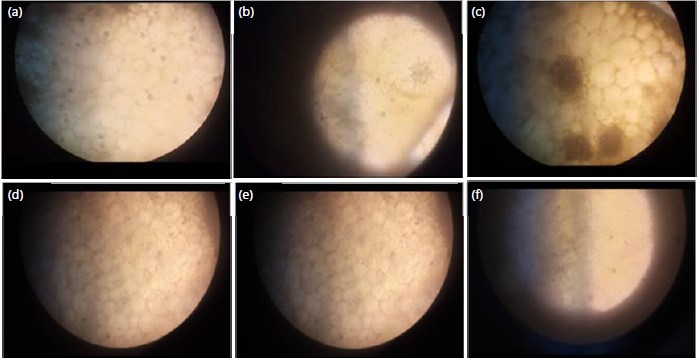
Stem histochemistry: The results of the stem histochemistry structures of the plants studied (Melissa officinalis, Peperomia pellucida and Portulaca oleracea) were presented in Fig. 3.
|
|
Melissa officinalis: There are calcium oxalate crystals in the form of crystal sand found in the cortex region (parenchyma cells) and the intercellular air spaces.
Portulaca oleraceae: There are calcium oxalate crystals in the form of druses in the parenchyma cells and intercellular air spaces. The druses are more numerous in the pith cavity than in the cortex region.
Peperomia pellucida
There were no calcium oxalate crystals.
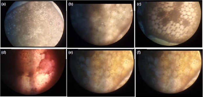
Petiole histochemistry: The results of the petiole histochemistry structures of the plants studied (Melissa officinalis, Peperomia pellucida and Portulaca oleracea) were presented in Fig. 4.
Melissa officinalis: There are calcium oxalate crystals in the form of druses in the intercellular air spaces and there is also crystal sand in the parenchyma cells and intercellular air spaces.
Portulaca oleracea: There are calcium oxalate crystals in the form of druses and crystal sand in the cortex and intercellular air spaces.
Peperomia pellucida: There are calcium oxalate crystals in the form of druses at the intercellular air spaces.
DISCUSSION
The anatomical results obtained from the observation of the stem of the 3 plants studied (Portulaca oleracea, Melissa officinalis and Peperomia pellucida) showed that they vary in the number of vascular bundles, layers of parenchyma and collenchyma cells. There were similarities in the pith cavity which is filled with parenchyma cells. The parenchyma cells have intercellular spaces within the cells. Its primary functions are photosynthesis, storage and buoyancy while the primary function of the collenchyma is providing support to withstand the forces of nature.
The results from the stem anatomy of M. officinalis showed that the stem is square shaped, consisting of a layer of the epidermis, followed by 2 to 3 layers of collenchyma and parenchyma cells which surround a layer of endodermis and pericycle. The vascular bundles are arranged in a ring, collateral and open and conform to the findings made by Nikitina et al.7, Shakeri et al.8 and Moradkhani et al.9.
Stem anatomical characteristics of P. pellucida were similar to the structures found in M. officinalis but differ in the collenchyma and parenchyma cells which are 3 to 4 layers and also lacking trichomes and this corresponds to the findings by da Silva et al.10, Budel and do Rocio Duarte11 and Camelo et al.12.
The results from the stem anatomical characters of P. oleracea also showed similarities to the structures in P. pellucida and M. officinalis but, the major differences were indicated in the number of layers of parenchyma cells which corresponds to the findings by Kumar et al.13 and Kumar et al.14.
The anatomical results gotten from the observation of the petiole of the 3 plants studied (Portulaca oleracea, Melissa officinalis and Peperomia pellucida) showed that there are similarities in their epidermal layer which is covered by a thin cuticle whose primary function is for protection against desiccation and external environmental stress. There are differences in the number and arrangement of vascular bundles as well as the layers of the parenchyma and collenchyma cells. This proves the fact that these plants are of different species. The results from the petiole anatomy of M. officinalis correspond to the findings by Kadam et al.15, Liu and Liu16.
The histochemical characteristics of the stem of the plants studied (Portulaca oleracea, Melissa officinalis and Peperomia pellucida) showed that there are calcium oxalate crystals found in two (Portulaca oleracea and Melissa officinalis) out of the 3 plants studied. There are no calcium oxalate crystals in Peperomia pellucida.
The histochemical characteristics of the petiole of the plants studied (Portulaca oleracea, Melissa officinalis and Peperomia pellucida) showed that the 3 plants studied possess calcium oxalate crystals and intercellular air spaces. The intercellular air spaces are important in the petiole for gas exchange and water transport.
The three plants studied in this research have high medicinal properties hence, most researchers based their studies on the medicinal, ethnobotanical, pharmacological and antioxidant properties, leading to very limited studies on their anatomy and histochemistry. Knowledge of the anatomical structures of these plants is very important as it will reduce the rate of adulteration when preparing the plant parts for medicinal purposes.
Results from the histochemical study showed that the plants contain calcium oxalate crystals which are protective in function and are a pointer to their proliferation as weeds. This will help in the production of an effective herbicide which will help to eliminate them since they are all invasive in nature.
CONCLUSION
As a result of this study, the anatomical features of plant materials have been established, which can be used in the identification and assessment of their authenticity, especially during preparations for medicinal purposes. Anatomical characteristics are essential since it portrays the physical appearance of the plants and also reflects the genetic makeup of the plant. This is taxonomically significant and knowledge of taxonomy is important for understanding the functioning of biodiversity and ecosystem. Further investigations in other areas such as chemotaxonomy and molecular genetics should be carried out to authenticate the classification of these plants into different species. The leaves of the three plants studied (Portulaca oleracea, Melissa officinalis and Peperomia pellucida) should also be given importance as it is greatly overlooked.
SIGNIFICANCE STATEMENT
There is a dearth of information on the anatomical and histochemical structures of the three plants studied: Melissa officinalis, Portulaca oleracea and Peperomia pellucida. This study will provide data on the histochemical structures of the plants studied: Melissa officinalis, Portulaca oleracea and Peperomia pellucida.
REFERENCES
- Ahn, K., 2017. The worldwide trend of using botanical drugs and strategies for developing global drugs. BMB Rep., 50: 111-116.
- Ward, S.M., R.D. Cousens, M.V. Bagavathiannan, J.N. Barney and H.J. Beckie et al., 2014. Agricultural weed research: A critique and two proposals. Weed Sci., 62: 672-678.
- Rao, V.S., 2000. Principles of Weed Science. 2nd Edn., CRC Press, Boca Raton, ISBN: 9780429081323, Pages: 564.
- Chandrasekaran, B., K. Annadurai and E. Somasundaram, 2010. A Textbook of Agronomy. 1st Edn., New Age International (P) Ltd., New Delhi, India, ISBN: 9788122427431, Pages: 835.
- Lyle, K.L., 2010. The Complete Guide to Edible Wild Plants, Mushrooms, Fruits, and Nuts. 2nd Edn., Globe Pequot Press, United States, ISBN 13: 978-0-7627-6303-0, Pages: 224.
- Scholey, A., A. Gibbs, C. Neale, N. Perry and A. Ossoukhova et al., 2014. Anti-stress effects of lemon balm-containing foods. Nutrients, 6: 4805-4821.
- Nikitina, A.S., L.A. Logvinenko, N.V. Nikitina and S.A. Nigaryan, 2018. Morphometric and histochemical research of Melissa officinalis L. herb from the collection of Nikitsky botanic garden. Pharm. Phamacol., 6: 504-534.
- Shakeri, A., A. Sahebkar and B. Javadi, 2016. Melissa officinalis L.-A review of its traditional uses, phytochemistry and pharmacology. J. Ethnopharmacol., 188: 204-228.
- Moradkhani, H., E. Sargsyan, H. Bibak, B. Naseri, M. Sadat-Hosseini, A. Fayazi-Barjin and H. Meftahizade, 2010. Melissa officinalis L., a valuable medicine plant: A review. J. Med. Plants Res., 4: 2753-2759.
- da Silva, R.M.F., G.C.R. de Medeiros, S.P.M. Costa, J.A. Gell, J.O.C.S. Júnio and P.J.R. Neto, 2014. Anatomical characterization and microchemistry of Peperomia pellucida (L.) H.B.K. (Piperaceae). Int. J. Pharm. Sci. Res., 5: 805-810.
- Budel, J.M. and M. do Rocio Duarte, 2010. Macro and microscopic characters of the aerial vegetative organs of Carqueja: Baccharis usterii Heering. Braz. Arch. Biol. Technol., 53: 123-131.
- Camelo, S.R.P., R.S. Costa, J.G. de Pontes Vieira, R.M. Ribeiro-Costa, W.L.R. Barbosa, F. Vasconcelos and J.O.C.S. Junior, 2012. Anatomical characterization and microchemistry of Vismia guianensis (Aubl.) Choisy (Clusiaceae) blade leaf. Int. J. Pharm. Sci. Res., 3: 1312-1317.
- Kumar, D., R. Kaur and Sonia, 2018. Morphological and anatomical studies of purslane (Portulaca oleracea) weed-an ethno medicinal plant. Int. J. Pharma Biol. Sci., 8: 1185-1189.
- Kumar, B.S.A., V. Prabhakarn, K. Lakshman, R. Nandeesh and P. Subramanyam et al., 2008. Pharmacognostical studies of Portulaca oleracea Linn. Braz. J. Pharmacogn., 18: 527-531.
- Kadam, V.B., S.S. Tambe, F. Sumia and R.K. Momin, 2013. Histochemical investigation of two medicinal plants in Maharashtra. J. Drug Delivery Ther., 3: 62-64.
- Liu, M.Q. and J.F. Liu, 2012. Structure and histochemistry of the glandular trichomes on the leaves of Isodon rubescens (Lamiaceae). Afr. J. Biotechnol., 11: 4069-4078.
How to Cite this paper?
APA-7 Style
Gospel,
A.M., Worlu,
F. (2023). Structural Characterization of Some Medicinal Weeds (Portulaca oleracea, Melissa officinalis and Peperomia pellucida) at Rivers State University. Trends in Agricultural Sciences, 2(3), 265-273. https://doi.org/10.17311/tas.2023.265.273
ACS Style
Gospel,
A.M.; Worlu,
F. Structural Characterization of Some Medicinal Weeds (Portulaca oleracea, Melissa officinalis and Peperomia pellucida) at Rivers State University. Trends Agric. Sci 2023, 2, 265-273. https://doi.org/10.17311/tas.2023.265.273
AMA Style
Gospel
AM, Worlu
F. Structural Characterization of Some Medicinal Weeds (Portulaca oleracea, Melissa officinalis and Peperomia pellucida) at Rivers State University. Trends in Agricultural Sciences. 2023; 2(3): 265-273. https://doi.org/10.17311/tas.2023.265.273
Chicago/Turabian Style
Gospel, Ajuru, Mercy, and Favour Worlu.
2023. "Structural Characterization of Some Medicinal Weeds (Portulaca oleracea, Melissa officinalis and Peperomia pellucida) at Rivers State University" Trends in Agricultural Sciences 2, no. 3: 265-273. https://doi.org/10.17311/tas.2023.265.273

This work is licensed under a Creative Commons Attribution 4.0 International License.