Effect of Secnidazole on the Disposition Kinetics of Diminazene Aceturate in Healthy Dogs
-
Ifeanyi G. Eke


Department of Veterinary Physiology and Pharmacology, Faculty of Veterinary Medicine, University of Nigeria, Nsukka, Enugu, Nigeria
Ukamaka U. Eze
Department of Veterinary Medicine, Faculty of Veterinary Medicine, University of Nigeria, Nsukka, Enugu, Nigeria
Ikenna O. Ezeh
Department of Veterinary Parasitology and Entomology, Faculty of Veterinary Medicine, University of Nigeria, Nsukka, Enugu, Nigeria
Terry A. Nzeakor
Department of Veterinary Parasitology and Entomology, Faculty of Veterinary Medicine, University of Nigeria, Nsukka, Enugu, Nigeria
Aruh O. Anaga
Department of Veterinary Physiology and Pharmacology, Faculty of Veterinary Medicine, University of Nigeria, Nsukka, Enugu, Nigeria
Patrick A. Onyeyili
Department of Veterinary Physiology and Pharmacology, Faculty of Veterinary Medicine, Federal University of Agriculture Makurdi, Benue, Nigeria
| Received 15 May, 2022 |
Accepted 08 Jul, 2022 |
Published 01 Oct, 2022 |
Background and Objective: Secnidazole (SEC) has been successfully combined with DA in the treatment of canine trypanosomosis without relapse parasitemia. However, the nature of the interaction between SEC and DA is not known. This study evaluated the influence of SEC on the disposition kinetics of DA in a healthy dog. Materials and Methods: Twelve dogs of both sexes were assigned to 2 groups A and B (n = 6). Group A was pre-treated with SEC (100 mg kg–1) 30 min before administration of DA (3.5 mg kg–1) IM. Group B received DA (3.5 mg kg–1) alone IM. Zero-time blood sample was obtained 15 min before DA administration and blood samples were obtained from each at 0.25, 0.5, 1, 2, 3, 6, 9, 12, 24, 36, 48, 60 and 72 hrs post-administration. Serum samples harvested were analysed for DA using the spectrophotometric method. Results: A double compartment model with a biphasic elimination best described the behaviour of DA in the two groups. Pre-treatment with SEC significantly (p<0.05) altered the Cmax,elimination rate constant, elimination half-life, total body clearance and area under the concentration-time curve of DA. Conclusion: Results concluded that the observed effects of SEC on the pharmacokinetic profile of DA may impact positively the efficacy of DA in trypanosome infected dogs.
| Copyright © 2022 Eke et al. This is an open-access article distributed under the Creative Commons Attribution License, which permits unrestricted use, distribution, and reproduction in any medium, provided the original work is properly cited. |
INTRODUCTION
Diminazene Aceturate (DA) is the most common agent used in the treatment of African animal trypanosomosis and babesiosis1. Canine trypanosomosis is marked by high morbidity, while poorly managed cases are usually fatal. Recurring infection, relapse parasitemia and drug resistance are the major causes of treatment failures2. Treatment failures due to drug resistance and relapse parasitemia and the unavailability of effective new trypanocides have encouraged the search for drug combinations in the treatment of trypanosomosis3. Amiodarone and itraconazole have been combined in the treatment of the chagas disease in dogs4. Eflornithine and nifurtimox are effective in the second stage of human African trypanosomosis5. However, toxicity concerns and ineffectiveness of some of these drugs have restricted the use of these drug combinations in clinical practice. The pharmacokinetics of DA has been explored in various animal species6-10. Although DA has been employed in the treatment of both canine trypanosomosis and babesiosis, only a few kinetic studies have been carried out with the drug in dogs. To the best of our knowledge, no kinetic work has been done on DA when combined with other drugs in dogs. Recently the antitrypanosomal effect of SEC in vitro and in vivo was reported11. Further studies have shown the efficacy of the combination therapy of SEC-DA in the canine model of trypanosomosis12. However, the nature of this synergistic effect of SEC-DA is not known.
The objective of this study was to determine the effect of SEC on the disposition kinetics of DA in healthy Nigerian indigenous breeds of dogs.
MATERIALS AND METHODS
Study area: The experimental animals were kept in the experimental kennels of the Department of Veterinary Physiology and Pharmacology Animal House. The pharmacokinetic studies were carried out at the World bank assisted by the Step B Drug Discovery Lab of the Department of Veterinary Physiology and Pharmacology University of Nigeria, Nsukka. The studies were carried out between February and May, 2016.
Twelve dogs of both sexes were used in the experiment. They were between 8-10 months old and were certified healthy. The dogs were kept in screened kennels in separate compartments. The dogs were fed with standard dry dog food (Dog and Co.® for puppies and junior. Adragna, Pet Food Via Porta Palermo, Italy). They were provided water ad libitum. Before the study fully commenced, they were clinically examined, wormed and blood samples collected and examined for the presence of trypanosomes and other blood parasites using Giemsa-stained blood smears. Four weeks of acclimatization were allowed before the studies commenced.
Ethical statement: This study was approved by the Experimental Animal Ethics Committee of the Faculty of Veterinary Medicine, University of Nigeria, Nsukka (Approval No: UNFVM/08/15/4). The protocol complied with the European Community Council Directive of November 24, 1986 (86/609/EEC) and the Federation of European Laboratory Animal Science Association.
Drugs:
| • | Secnidazole (Secwid®) May and Baker Nig. Ltd., Lagos Nigeria |
| • | Diminazene aceturate (Veriben®) Ceva Sante Animale, Libourne Cedex, France |
| • | Pure diminazene aceturate for preparation of standard (Cayman chemical, Michigan, U.S.A) |
Chemicals: Trichloroacetic acid (TCA) (Sigma-Aldrich, Darmstadt Germany), N-HCl (New Bombay Acid and Chemical Company, Bombay, India), sodium nitrite (Airedale Chemical Company Ltd, West Yorkshire, UK), Ammonium sulphamate (Sigma-Aldrich, Darmstadt Germany). Alpha-naptylethylenediamine (Sigma-Aldrich, Damstadt Germany).
Experimental animals and treatments: The dogs were randomly assigned to 2 groups (n = 6). The first group was treated with 100 mg kg–1 SEC orally followed by 3.5 mg kg–1 DA IM 30 min later. The DA alone 3.5 mg kg–1 IM was given to the second group.
Diminazene aceturate 7% solution was administered IM to the dogs in the left gluteal muscle, while blood samples were collected from the right cephalic vein.
Sample collection: About 15 min before drug administration zero-time (0 hr) samples were collected from each dog. After drug administration, blood samples were collected at 0.25, 0.5, 1, 2, 3, 6, 9, 12, 24, 36, 48, 60 and 72 hrs. The samples were centrifuged at 4000 rpm immediately after collection for 5 min, to obtain the serum. The serum samples were stored frozen until analysed.
Preparation of DA standard: Pure diminazene aceturate (2 mg mL–1) solution was prepared. From this stock solution, 20 μg mL–1 was prepared. Two millilitres of this solution were further diluted with the serum to obtain 4, 2, 1, 0.5, 0.25, 0.125 and 0.0625 μg mL–1. The optical density readings of the various concentrations were obtained using a double beam UV-spectrophotometer (Jenway, Staffordshire, UK). The standard curve was generated and the standard equation was derived using a Microsoft excel package.
Analysis of DA in serum: Total DA was determined using the spectrophotometric method13. The procedure is based on the hydrolysis of DA and coupling of the diazonium salt of the drug with N-1-naphthyl ethylenediamine to form a pink colour. Treatment of DA with HCl and sodium nitrite solutions results in the formation of diazonium salt from the DA present. The excess nitrous acid was destroyed using ammonium sulphamate, leaving the diazonium salt to be coupled with N-1-naphthyl ethylenediamine. The colour change was measured with a UV spectrophotometer.
Protocol: To 1 mL of 10% TCA, 1 mL of serum was added. The sample was left to stand for 15 min after thorough shaking. The sample was centrifuged for 10 min at 4000 rpm. Clear supernatant solution 1 mL was added to 1 mL of 1N-HCl in a test tube. The mixture was diazotized with 0.2 mL of 0.5% sodium nitrite solution. One millilitre of 1% ammonium sulphamate solution was added to the mixture after 3 min and the mixture was shaken well. One millilitre of 1 mL of 0.2% alpha-naphtyl ethylene diamine solution was added after an additional 3 min and the developed colour was measured at 540 nm using a double beam UV-spectrophotometer (Jenway, Staffordshire, UK).
Calculation of DA concentrations: The concentrations of DA in the serum were calculated from the standard curve which was obtained from the optical density readings of the standard.
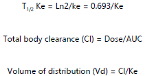
Calculation of pharmacokinetic parameters: Pharmacokinetic analysis of data obtained was performed using a mean value by standard procedure14-16. The following pharmacokinetic parameters were determined.Where:
| D | = |
Dose of DA administered |
| Cl | = |
Total body clearance |
Elimination rate constant:
Where
| C1 T1 | = |
First drug serum concentration and time pair |
| C2 T2 | = |
Second drug serum concentration and time pair |
Elimination half-life:
 |
| • | Absorption rate constant (Ka): Derived by method of Residuals16 |
| • | Absorption half-life (T1/2Ka) = Ln2/Ka = 0.693/Ka |
| • | Tmax = Time of maximum drug concentration in the serum |
| • | Cmax = Maximum drug concentration in the serum |
Statistical analysis: The results were presented as Mean±SEM. Data obtained were analysed using an independent sample T-test. Significance was accepted at p<0.05.
RESULTS
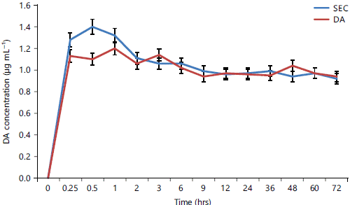
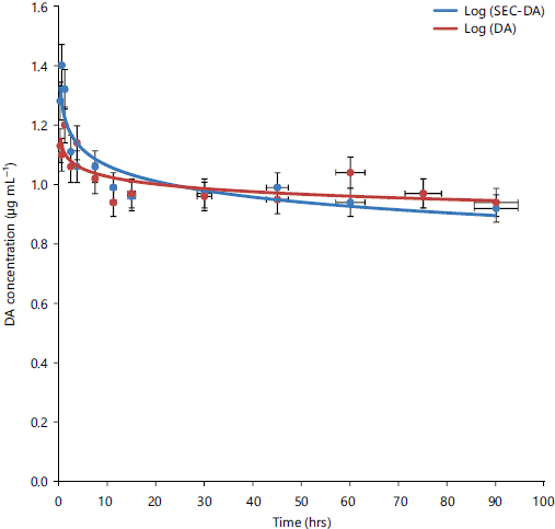
Figure 1 showed that DA concentration was significantly (p<0.05) higher in SEC pre-treated dogs at 0.25 and 0.5 hr. There was no significant variation in the DA concentration afterwards. The DA was rapidly absorbed after intramuscular administration in the two groups of dogs as shown in Fig. 2. A double compartment model, characterized by biphasic elimination best described the DA data presented in the figure. The mean peak serum concentration of 1.40±0.2 μg mL–1 of DA achieved at 0.5 hr in SEC pre-treated dogs was significantly (p<0.05) higher than that obtained in the DA alone treated dogs (1.2±0.05) at 1 hr. Pre-treatment with SEC significantly (p<0.05) increased the Ke of DA when compared with DA alone. There was a significantly (p<0.05) lower T1/2Ke of DA in the SEC pre-treated dogs compared to DA alone. There was significantly (p<0.05) higher Cl and AUC of DA in SEC pre-treated dogs compared to DA alone. There was no significant (p>0.05) variation in the Ka, T1/2Ka and Vd of DA in dogs in both treatment groups (Table 1).
|
|
| Table 1: | Pharmacokinetic parameters of DA in dogs following administration of SEC-DA and DA alone | |||
| Pharmacokinetic parameters | SEC-DA (n = 6)a |
DA (n = 6)a |
| Tmax (hrs) | 0.50±0.00 |
1.00±0.00 |
| Cmax (μg mL–1) | 1.40±0.20 |
1.20±0.05b |
| Elimination rate constant (Ke) (hrs–1) | 0.04±0.01 |
0.01±0.03b |
| Elimination half-life (T1/2Ke) (hrs) | 18.97±3.40 |
64.00±7.9b |
| Absorption rate constant (Ka) (hrs–1) | 7.06±0.46 |
6.93±0.37 |
| Absorption half-life (T1/2Ka) (hrs) | 0.10±0.01 |
0.10±0.01 |
| Volume of distribution (Vd) (L kg–1) | 3.03±0.12 |
3.25±0.07 |
| Total body clearance (Cl) (L hrs–1) | 0.12±0.01 |
0.05±0.01b |
| Area under the curve (AUC) (hrs μg mL–1) | 30.27±4.40 |
72.20±2.2b |
| aData represents Mean±SEM, bData significantly different from those of SEC-DA treatment (p<0.05) | ||
DISCUSSION
The data obtained from this study indicated that DA was absorbed from the IM route. The disposition kinetics of DA in the two treatment groups were similar. However, pre-treatment of dogs with SEC increased the Cmax and decreased the Tmax of DA. Also, pre-treatment of dogs with SEC increased the elimination rate constant and total body clearance and decreased the elimination half-life and AUC. A double compartment model, characterized by biphasic elimination best described the disposition characteristics of DA in both treatment groups.
The absorption rate constant is the fractional rate of drug absorption from the site of administration into the systemic circulation14. Pre-treatment with SEC had no significant effect on the absorption of DA from the IM injection site. However, the absorption of DA from the injection site into the blood was very rapid (0.1 hr) and was similar to the findings of previous researchers10. A biphasic elimination of DA was observed for both treatments with a more prolonged elimination in the DA alone treated dogs. This finding agrees with the findings of Miler et al.10 who reported that DA has rapid and slow terminal elimination phases.
An elimination rate constant of 0.04 per hr was determined for dogs pre-treated with SEC as against 0.01 per hr for dogs treated with DA alone. The elimination half-life was also shorter for dogs pre-treated with SEC (18.97 hrs) against 64.00 hrs for dogs treated with DA. The reason for this was not clear, but it could be related to increased clearance of the serum of DA into organs and tissues. A previous study showed that SEC pre-treated dogs have higher DA concentrations in the liver, kidney, brain and skeletal muscles13. This has chemotherapeutic implications in the treatment of animal trypanosomosis. Relapse parasitemia is due to sequestration of trypanosomes in sites like the brain where DA accumulation is below therapeutic levels17,18. Increased accumulation of DA in these sites may lead to the elimination of relapse parasitemia13. This may explain the reported greater efficacy of SEC-DA over DA alone in Trypanosoma brucei brucei infected dogs12. The Volume of Distribution (Vd) is a factor that relates the amount of drug in the body to the concentration of drug in the serum14. In the current study, pre-treatment of dogs with SEC did not significantly alter the Vd of DA. Dogs pre-treated with SEC had a Vd of 3.03 L kg–1 against 3.25 L kg–1 for dogs treated with DA alone. However, the results obtained showed that DA was distributed at least into the interstitial fluid spaces and tissues in both groups19. Clearance reflects the elimination of drugs from the body15. Thus, clearance is the coefficient of proportionality between serum drug concentration and elimination. Total body clearance of DA was significantly (p<0.05) higher (0.12 L hr–1) in dogs pretreated with SEC compared with 0.05 L hr–1 for dogs treated with DA alone. This explained the higher rate of elimination of DA in the SEC pre-treated dogs. This will suggest lower toxicity20,21. The Area Under the Concentration-Time Curve (AUC) is the integral of the serum drug concentration-time curve15. The AUC reflects the actual body exposure to the drug after administration of a dose of the drug15. It is dependent on the rate of elimination and dose administered and inversely proportional to the drug clearance22. In the current study, pre-treatment with SEC decreased the AUC (30.27 hrs μg mL–1) against 72.20 hrs μg mL–1 for DA alone. This suggested that the body exposure to the drug was lower in SEC pre-treated dogs than in DA because of higher clearance and faster decline in the serum drug concentration. In another study, Baker et al.23 showed that AUC was directly correlated to neutropenia in 640 patients receiving docetaxel. Clinically, the implication of decreased AUC in SEC pre-treated dogs could be reduced risk of DA toxicity or no toxicity at all24. One of the important limitations of this study is that it was conducted in healthy dogs and under controlled experimental conditions. What happens in infected or natural field infection remains speculative. Thus, we, therefore, suggest that this work be repeated in trypanosome infected dogs to get a complete overview of the pharmacokinetic interaction of SEC and DA.
CONCLUSION
The SEC altered some pharmacokinetic profiles and elimination patterns of DA in treated dogs. The shortened elimination half-life, increased clearance and lower AUC of DA observed in SEC pre-treated dogs may be evidence that SEC pre-treatment could decrease DA toxicity in treated dogs. This is of clinical significance since DA toxicity is a huge concern in chemotherapy of canine trypanosomosis. These effects of SEC may impact positively on the efficacy of DA in the treatment of canine trypanosomosis.
SIGNIFICANCE STATEMENT
Previous studies had demonstrated the efficacy of combination therapy of SEC-DA in Trypanosoma brucei brucei infected dogs. However, the possible mechanism of synergy between SEC and DA was speculative. The current study provides substantial evidence of pharmacokinetic interaction between the two combined agents. This study is one of the very few pharmacokinetic studies involving the combination of other agents with diminazene aceturate and the first of such studies in dogs.
REFERENCES
- Cossic, B.G.A., B. Adjahoutonon, P. Gloaguen, G.L. Dibanganga and G. Maganga et al., 2017. Trypanosomiasis challenge estimation using the diminazene aceturate (Berenil) index in Zebu in Gabon. Trop. Anim. Health Prod., 49: 619-624.
- Giordani, F., L.J. Morrison, T.G. Rowan, H.P. de Koning and M.P. Barrett, 2016. The animal trypanosomiases and their chemotherapy: A review. Parasitology, 143: 1862-1889.
- Suganuma, K., D.D. N’Da, K.I. Watanabe, Y. Tanaka and E. Mossaad et al., 2022. Therapeutic efficacy of orally administered nitrofurantoin against animal African trypanosomosis caused by Trypanosoma congolense infection. Pathogens.
- Madigan, R., S. Majoy, K. Ritter, J.L. Concepción and M.E. Márquez et al., 2019. Investigation of a combination of amiodarone and itraconazole for treatment of American trypanosomiasis (Chagas disease) in dogs. J. Am. Vet. Med. Assoc., 255: 317-329.
- Priotto, G., S. Kasparian, W. Mutombo, D. Ngouama and S. Ghorashian et al., 2009. Nifurtimox-eflornithine combination therapy for second-stage African Trypanosoma brucei gambiense trypanosomiasis: A multicentre, randomised, phase III, non-inferiority trial. Lancet, 374: 56-64.
- Pandey, H.K., K.K. Singh, B.K. Roy and S. Kumari, 2010. Pharmacokinetics of diminazene aceturate in buffalo calves. J. Bioanalysis Biomed., 2: 13-16.
- El Banna, H.A., K.A. El-Sooud and G.A. Soliman, 1999. Comparative pharmacokinetics of diminazene in lactating goats and sheep. J. Vet. Med. Ser. A, 46: 49-58.
- Lewis, K.M., L.A. Cohn, A.J. Birkenheuer and M.G. Papich, 2012. Pharmacokinetics of diminazene diaceturate in healthy cats. J. Vet. Pharmacol. Ther., 35: 608-610.
- Staroverov, S.A., V.A. Sidorkin, A.S. Fomin, S.Y. Shchyogolev and L.A. Dykman, 2011. Biodynamic parameters of micellar diminazene in sheep erythrocytes and blood plasma. J. Vet. Sci., 12: 303-307.
- Miller, D.M., G.E. Swan, R.G. Lobetti and L.S. Jacobson, 2005. The pharmacokinetics of diminazene aceturate after intramuscular administration in healthy dogs. J. South Afr. Vet. Assoc., 76: 146-150.
- Eke, I.G., I.O. Eze, T.A. Ezeudu, U.U. Eze, A.O. Anaga and P.A. Onyeyili, 2017. Anti-trypanosomal activity of secnidazole in vitro and in vivo. Trop. J. Pharm. Res., 16: 535-541.
- Eke, I.G., I.O. Ezeh, T.A. Ezeudu, U.U. Eze, A.O. Anaga and P.A. Onyeyili, 2020. Efficacy of secnidazole-diminazene aceturate combination therapy in the late treatment of Trypanosoma brucei brucei infection in dogs. Braz. J. Pharm. Sci.
- Eke, I.G., U.U. Eze, T.A. Ezeudu, I.O. Ezeh, A.O. Anaga and P.A. Onyeyili, 2017. Diminazene aceturate residues in tissues of dogs treated with secnidazole-diminazene aceturate combination and with diminazene aceturate alone. Sokoto J. Vet. Sci., 15: 16-20.
- Bauer, L., 2008. Applied Clinical Pharmacokinetics. 2nd Edn., McGraw-Hill Education, New York, ISBN: 0071476288, Pages: 826.
- Gustafson, D.L. and E.L. Bradshaw-Pierce, 2010. Fundamental Concepts in Clinical Pharmacology. In: Principles of Anticancer Drug Development, Garrett-Mayer, E. (Ed.), Springer, New York, ISBN 978-1-4419-7357-3 pp: 37–62.
- Odeh, A.J., O.P. Azubuike, S.A. Saganuwan and B.J. Aondohulugh, 2019. Effect of age on the pharmacokinetics of sulphadimidine in West African Dwarf (WAD) goats following a single intramuscular administration. GSC Biol. Pharm. Sci., 9: 44-49.
- Masocha, W., M.E. Rottenberg and K. Kristensson, 2007. Migration of African trypanosomes across the blood-brain barrier. Physiol. Behav., 92: 110-114.
- Emiru, A.Y., E. Makonnen, F. Regassa, F. Regassa and T.B. Tufa, 2021. Antitrypanosomal activity of hydromethanol extract of leaves of Cymbopogon citratus and seeds of Lepidium sativum: In-vivo mice model. BMC Complementary Med. Ther.,
- Bhosle, V.K., G. Altit, J. Autmizguine and S. Chemtob, 2017. Basic Pharmacologic Principles. In: Fetal and Neonatal Physiology, Polin, R.A., S.H. Abman, D.H. Rowitch, W.E. Benitz and W.W. Fox (Eds.), Elsevier, Amsterdam, ISBN: 9780323352147, pp: 187-201.
- Hristova, V. and W. Clarke, 2016. Therapeutic Drug Monitoring in Obese Patients. In: Clinical Challenges in Therapeutic Drug Monitoring, Clarke, W. and A. Dasgupta (Eds.), Elsevier, Amsterdam, ISBN: 978-0-12-802025-8, pp: 231-243.
- Yamazaki, H., 2017. Drug-Metabolizing Enzyme Systems I. In: Comprehensive Medicinal Chemistry III, Chackalamannil, S., D. Rotella and S.E. Ward (Eds.), Elsevier, Amsterdam, ISBN: 978-0-12-803201-5, pp: 45-50.
- Sahota, T., M. Danhof and O.D. Pasqua, 2015. The impact of composite AUC estimates on the prediction of systemic exposure in toxicology experiments. J. Pharmacokinet. Pharmacodynamics, 42: 251-261.
- Baker, S.D., J. Li, A.J.T. Tije, W.D. Figg, W. Graveland, J. Verweij and A. Sparreboom, 2005. Relationship of systemic exposure to unbound docetaxel and neutropenia. Clin. Pharmacol. Ther., 77: 43-53.
- Sun, N., B. Shen, J. Zhu, X. Zhang and H. Zhu et al., 2020. Clinical application of the AUC-guided dosage adjustment of docetaxel-based chemotherapy for patients with solid tumours: A single centre, prospective and randomised control study. J. Transl. Med.
How to Cite this paper?
APA-7 Style
Eke,
I.G., Eze,
U.U., Ezeh,
I.O., Nzeakor,
T.A., Anaga,
A.O., Onyeyili,
P.A. (2022). Effect of Secnidazole on the Disposition Kinetics of Diminazene Aceturate in Healthy Dogs. Trends in Agricultural Sciences, 1(2), 95-102. https://doi.org/10.17311/tas.2022.95.102
ACS Style
Eke,
I.G.; Eze,
U.U.; Ezeh,
I.O.; Nzeakor,
T.A.; Anaga,
A.O.; Onyeyili,
P.A. Effect of Secnidazole on the Disposition Kinetics of Diminazene Aceturate in Healthy Dogs. Trends Agric. Sci 2022, 1, 95-102. https://doi.org/10.17311/tas.2022.95.102
AMA Style
Eke
IG, Eze
UU, Ezeh
IO, Nzeakor
TA, Anaga
AO, Onyeyili
PA. Effect of Secnidazole on the Disposition Kinetics of Diminazene Aceturate in Healthy Dogs. Trends in Agricultural Sciences. 2022; 1(2): 95-102. https://doi.org/10.17311/tas.2022.95.102
Chicago/Turabian Style
Eke, Ifeanyi, G., Ukamaka U. Eze, Ikenna O. Ezeh, Terry A. Nzeakor, Aruh O. Anaga, and Patrick A. Onyeyili.
2022. "Effect of Secnidazole on the Disposition Kinetics of Diminazene Aceturate in Healthy Dogs" Trends in Agricultural Sciences 1, no. 2: 95-102. https://doi.org/10.17311/tas.2022.95.102

This work is licensed under a Creative Commons Attribution 4.0 International License.